Figure 3.
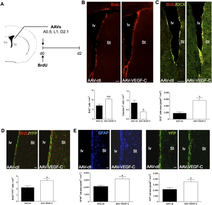
In vivo VEGF-C overexpression. (A) Adult mice are infected with either AAV-VEGF-C or control AAVs (AAV-ctl: AAV-EGFP and AAV-HSA) in the vicinity of the SVZ (coordinates from the bregma: anterior, 0.5; lateral, 1.0; depth, 2.1; n = 7 animals per group) and injected intraperitoneally with BrdU the same day. (B) Coronal brain cryosections from animals sacrificed at day 2 post-infection are analyzed for the number of BrdU cells (red) in the SVZ. Both the dorsolateral and ventral regions along the striatal wall show more BrdU+ cells in AAV-VEGF-C-treated animals as compared with AAV-ctl. Histograms indicate the number of newborn BrdU+ cells (bottom left) and activated Caspase-3+ cells (bottom right) in the SVZ. AAV-VEGF-C promotes the production of newborn cells and inhibits PCD in SVZ cells. (C) Coronal cryosections of the periventricular zone immunolabeled with anti-DCX Ab (green). In the striatal SVZ, DCX labeling reflects neuroblast production. AAV-VEGF-C induces a significant expansion of DCX expression in the SVZ compared with controls. (D,E) Coronal brain vibratome sections from Vegfr3:YFP adult mice infected with either AAV-VEGF-C or AAV-ctl and sacrificed at day 2. Dividing VEGFR-3-expressing cells (BrdU+YFP+) are increased in AAV-VEGF-C-treated animals (D), while the subventricular expressions of GFAP and YFP are also enhanced (E), as compared with controls.(St) Striatum. Bars: B,C, 100 μm; D,E, 20 μm. Error bars indicate SEM. (***) P < 0.001, Student's t-test.

