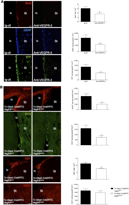
Figure 6.
(A) Function-blocking Abs against VEGFR-3 (31-C1: anti-VEGFR-3) or rat IgG2a (Ig-ctl, control) were delivered above the lv of adult Vegfr3:YFP mice with an Alzet miniosmotic pump for 6 d (coordinates from the bregma: anterior, 0.5; lateral, 1.0; depth, 0). Coronal brain vibratome sections are stained with specific markers as indicated (left panels), and histograms show the corresponding cell quantifications (right panels). Anti-VEGFR-3-treated mice display few BrdU+ cells and reduced expression patterns for GFAP and YFP in the SVZ, as compared with control animals. (B) Tx-induced deletion of Vegfr3 in subventricular astroglial cells in GlastCreERT2:Cre, Vegfr3lox/lox mice. Coronal cryosection of the adult lv walls showing reduction in the expression of GFAP and DCX, and in the number of dividing KI67+ cells. No changes were observed in the pattern of CD31+ endothelial cells. Quantifications confirm a phenotype similar to the one observed in Brn4:Cre, Vegfr3lox/lox mice. Bars: A, 20 μm; B, 50 μm. n = 3–4 animals per group. Error bars indicate SEM. (*) P < 0.05; (**) P ≤ 0.005, Student's t-test.

