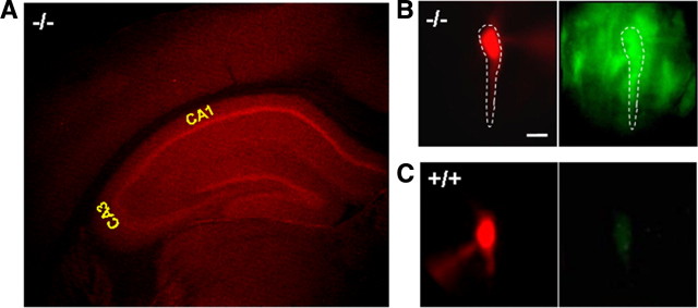
Figure 1.
Expression of Zip-1,3 Zn transporters in the hippocampus. A, Immunofluorescence staining of anti-GFP-Alexa 594 (Texas Red) in a coronal section of Zip-1,3-null mice labeled with targeted EGFP reporter. Intense signal indicates strong expression of Zip-1 and Zip-3 in the hippocampal pyramidal (CA1, CA3) and dentate granule cell layers with low levels in overlying neocortex. B, C, High-magnification EGFP fluorescence images taken from Zip-1,3 −/− (B) and +/+ (C) brain slices. Scale bar, 10 μm. To verify EGFP staining in identified CA1 neurons, Alexa dye was injected through a patch-clamp electrode and visualized with filtering for Alexa (B, left), then EGFP (B, right). In the mutant (B), fluorescent Alexa pyramidal cell (dotted outline) colabels EGFP-filled cells. In the wild-type cell (C), no EGFP is present (C, right). To highlight the somatic GFP fluorescence, the EGFP fluorescence intensity in the dendritic region was subtracted from the raw image data. To compare the EGFP fluorescence intensity between two genotypes, the EGFP image from the +/+ group is normalized to the one taken from the −/− group.

