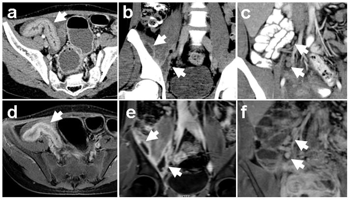
Figure 3.

MRI detection of IBD imaging features. Corresponding image pairs from contrast enhanced CT (top row) and MR enterography (bottom row) studies on the same patients with known IBD demonstrate bowel wall thickening (a, d), intramuscular abscesses (b, e), and mesenteric lymphadenopathy (c, f). Arrows indicate the abnormalities.
