
Figure 4. Ibuprofen treatment relieves IκBα-mediated repression of NF-κB-p65 and induces its phosphorylation and nuclear localization.
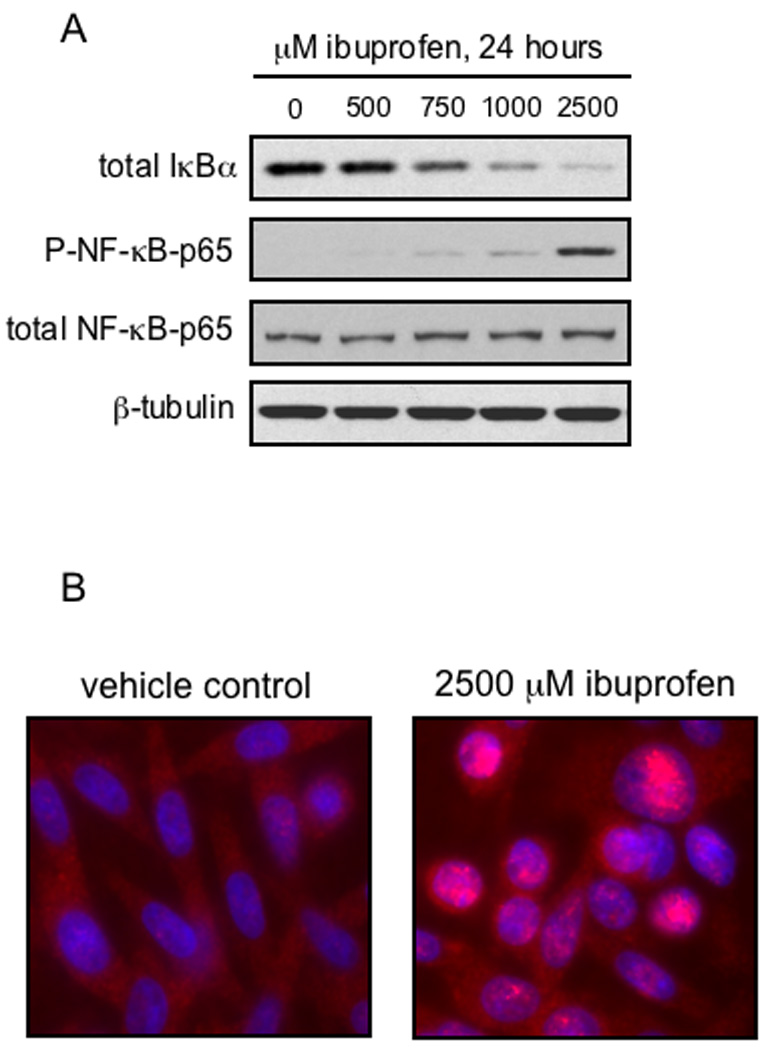
A, SW480 cells were treated for 24 hours with increasing doses of S-ibuprofen and protein lysates were collected as described under Materials and methods. Proteins were resolved by SDS-PAGE and blotted for IκBα, phospho-NF-κB-p65 (serine-536) and β-tubulin (loading control). The phospho-NF-κB-p65 blot was stripped and blotted for total NF-κB-p65. B, SW480 cells were grown overnight on cover-slips and then treated for 24 hours with either vehicle control or 2500 µM S-ibuprofen. Immunofluorescence microscopy was performed to visualize NF-κB-p65 (Alexa Fluor 568, red). Nuclei were counterstained with DAPI (blue). Localization of NF-κB-p65 in the nucleus is represented by magenta staining after merging of the two images.