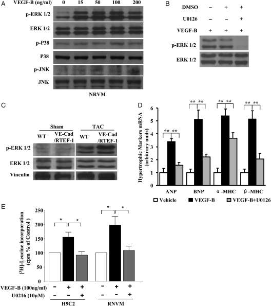
Figure 6.
VEGF-B induced cardiac hypertrophy via phosphorylation of ERK1/2. (A) Western blots of ERK1/2, p38, JNK, and their phosphorylated forms in NRVMs incubated with 0, 15, 50, 100, or 200 ng/mL of recombinant hrVEGF-B167 for 15 min. (B) Western blots of ERK1/2 and phosphorylated ERK1/2 in NRVMs incubated with 100 ng/mL of rhVEGF-B167, rhVEGF-B167 with DMSO, or rhVEGF-B167 plus 10 μM U0126 for 15 min. (C) Western blots of ERK1/2 and phosphorylated ERK1/2 in heart samples from VE-Cad/RTEF-1 mice or WT mice after sham treatment or after TAC. (D) qPCR showing expression of ANP, BNP, α-MHC, and β-MHC by H9C2 cells treated with 100 ng/mL of rhVEGF-B167 or rhVEGF-B167 plus 10 μM U0126. (E) 3H-Leucine incorporation assay indicates the level of protein synthesis in H9C2 and RNCM cultures after incubation of VEGF-B with and without ERK1/2 inhibitor U0216. Bars represent radioactivity of incorporated 3H-leucine (cpm). The data are expressed as the means ± SEM of three independent experiments. *P < 0.05 and **P < 0.01.

