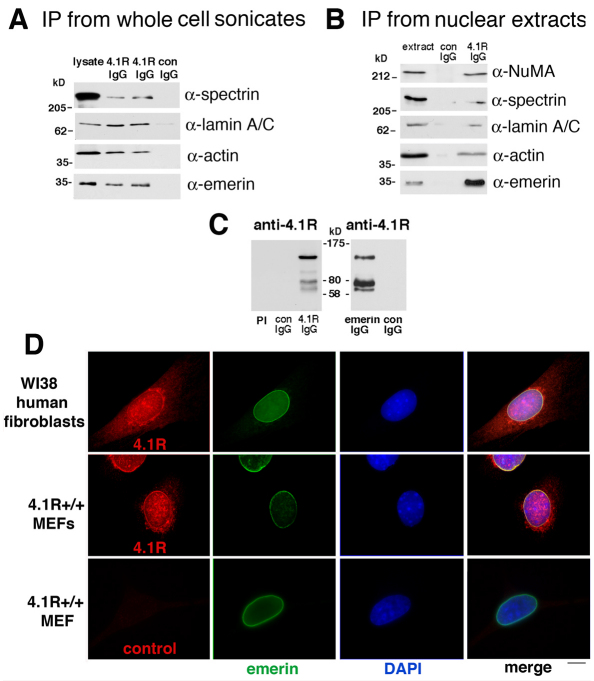
Fig. 1.
4.1R and emerin co-immunoprecipitate and partially colocalize in human and murine fibroblasts. (A) Immunoprecipitation (IP) from HeLa whole cell sonicates. Clarified whole cell sonicates were prepared, immunoprecipitated with anti-4.1R antibody, and eluted proteins separated by SDS-PAGE were subjected to western blotting as described in the Materials and Methods. Emerin, actin, lamin A/C and αII-spectrin were detected using the antibodies indicated in lysate and immune duplicate samples (4.1R IgG) but not in a non-immune sample (con IgG). (B) Analysis of immunoprecipitates from HeLa nuclear extract. HeLa nuclear extract was incubated with antibodies, immunoprecipitated with anti-4.1R antibody, and eluted proteins separated by SDS-PAGE were subjected to western blotting using the antibodies indicated on the right. In 4.1R IgG immunoprecipitates (right-hand lane) a strong 34 kDa emerin band was detected. 4.1R immunoprecipitates also contained actin, lamin A/C, αII-spectrin and NuMA. These proteins were not detected in control IgG precipitates (center lane). (C) Western blot probed with anti-4.1R antibody showing 4.1R bands migrating at ~135, 105, 80 and 62 kDa detected specifically in 4.1R IgG immunoprecipitates but not in the pre-immune (PI) or control (con) lanes. The right-hand panel shows a western blot demonstrating that 4.1R bands at ~135, 80, and 62 kDa were present in anti-emerin antibody immunoprecipitates but not in those with control IgG. (D) Partial colocalization of 4.1R epitopes (red) with emerin epitopes (green) in human and murine fibroblasts. Fixed WI38 fibroblasts and wild-type MEFs were probed by double-label indirect immunofluorescence. The nuclear envelope protein emerin was detected at the periphery of the nucleus stained by DAPI. 4.1R epitopes were detected in the nucleus, cytoplasm and in the region of the nuclear envelope, partially coincident with emerin signals (yellow coloration in the ‘merge’ image). Signals for an equal amount of control anti-rabbit IgG antibody staining (bottom left panel) were not above background levels of other controls in which primary rabbit antibody was omitted but murine anti-emerin and both fluorescent secondaries were included. Scale bar: 6 μm.

