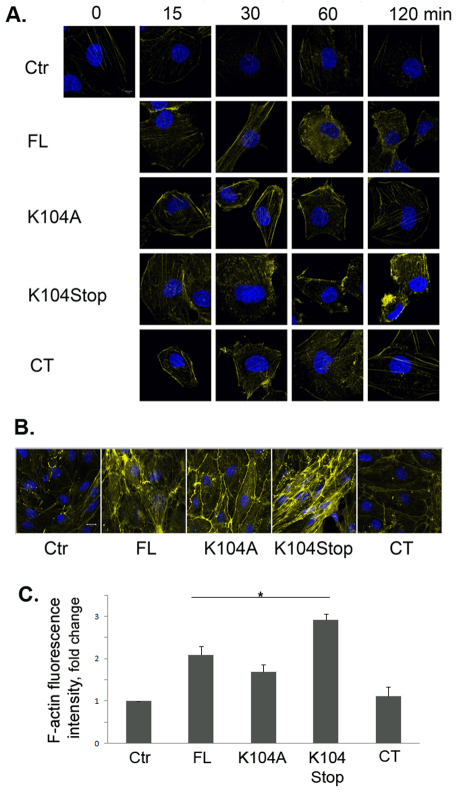
Fig. 5.
Truncated MCP1 promotes reorganization of the actin cytoskeleton. (A) HBMECs were treated with saline (Ctr) or the indicated form of MCP1 (100 nM) for different times, and the actin cytoskeleton was visualized using Alexa-Fluor-647–phalloidin. (B) Primary mouse BMECs treated with the indicated form of MCP1 (100 nM) for 2 hours were immunostained to visualize the actin cytoskeleton. (C) Quantification of F-actin fluorescence intensity. Values are mean+s.d. (n=9). *P<0.05 (analyzed using Student's t-tests).

