Fig. 2.
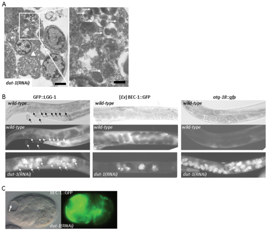
Depletion of DUT-1 or dUTPase in C. elegans causes a massive upregulation of autophagy genes. (A) Transmission electron microscopic image showing a cell (arrowhead) with extremely high autophagic activity in a dut-1(RNAi) embryo. The left panel displays an enlargement of the same cell with autophagic elements in various stages of the degradation process (arrows). Note that this cell also shows apoptotic features such as chromatin condensation. Scale bars: 1 μm (left) and 0.25 μm (right). (B) dUTPase deficiency leads to a massive upregulation of three autophagic markers, GFP::LGG-1, BEC-1::GFP and atg-18::gfp (Aladzsity et al., 2007; Sigmond et al., 2008). In wild-type embryos, these GFP-labeled reporters show a faint expression, which manifests as dark balls within the hermaphrodites on the fluorescence pictures (middle panels). The corresponding differential interference contrast (DIC) pictures are in the upper panels. Reporter expression in the dut-1(RNAi) background is shown in the bottom panels: dut-1(RNAi) embryos expressing the reporters are seen as brightly fluorescent balls within the hermaphrodites. Intense expression of BEC-1 is only restricted to several dut-1(RNAi) embryos (white balls), which is due to the mosaicism of the non-integrated BEC-1 reporter. Arrows indicate embryos inside a hermaphrodite transgenic for GFP::LGG-1. (C) Excessive accumulation of a functional BEC-1::GFP reporter in a dut-1(RNAi) embryo is associated with tissue atrophy. The arrow indicates a necrotic area. Left, DIC image; right, the corresponding fluorescent image.

