Figure 9.
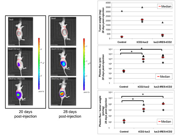
In vivo imaging of bioluminescent tumors containing two types of luciferase expression constructs. Stably transfected Daudi cells (6.106 cells) were injected subcutaneously into the flank of nude mice forming visible tumors on average two weeks later. The resulting tumors were imaged after intra-peritoneal injection of the D-luciferin reagent using an IVIS 50 (Xenogen). Three series of three mice were imaged 20 and 28 days following injection of wild-type Daudi cells (used as controls, upper line of the left panel), Daudi cells transfected with the monocistronic luciferase construct (medium line) or the bicistronic construct (bottom line). At day 28, tumors were collected and weighted just after the imaging procedure. In the right panel, are plotted tumor weights (upper graph), total photon flux (medium graph) and the ratios of photon flux to tumor weights (bottom graph). The stars indicate a statistical difference from controls (Kruskal-Wallis one-way analysis, p < 0.06). Although in this experiment, injection of Daudi cells containing the tCD2-luc2 construct resulted in bigger tumors than the wild-type Daudi cells, this effect was not observed in another experiment (data not shown). These data as well as the results presented in this figure and in Figure 8 confirm that the tCD2-luc2 protein does not prevent cell growth in vivo as well as in vitro.

