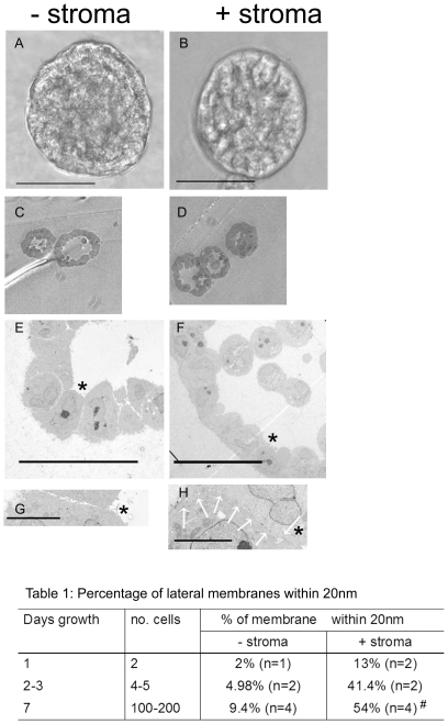
Figure 1. Stromal co-culture increases lateral epithelial cell adhesion.
(A and B). Phase images of 3D BPH-1 acini grown with (B) and without stroma (A) for 8 days, bars = 50 µm. Thick sections (C,D) and TEM (E,F) of mid-sections through BPH-1 acini, grown with (D,F) and without stroma (C,E). Bar = 20 (E) or 50 µm (F). (G and H). High magnification TEM images of the junctions marked by the asterisks in (E) and (F). The lateral cell-cell contact shown in (H) is highlighted by white arrows, bars = 5 µm. Images are representative of three spheroids analysed per experiment. The values in table 1 have standard deviations of ±15%, # = p<0.018.

