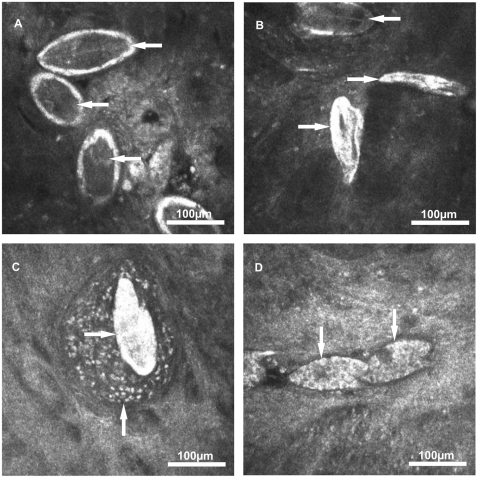
Figure 2. Eggs of S. mansoni visualised within the colon mucosa by image modality 1.
(A) Viable mature eggs containing dark coloured and fully developed miracidia (←). (B) Dead eggs without any content (→) and a mature egg containing a dark coloured miracidium (←). (C) Dead egg (→) with surrounding granulomatous tissue (↑). (D) Viable immature eggs of developmental stage 1 (↓) containing various vitelline cells.

