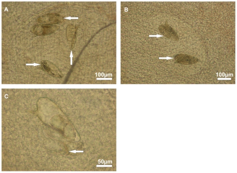
Figure 4. Eggs of S. mansoni visualised within a colon biopsy crush preparation by bright-field microscopy.
(A) Viable mature eggs containing fully developed miracidia (←), an empty egg shell (↑) of a previously hatched miracidium and a dead egg with diffuse contents (→). (B) Dead eggs without dark and granular contents (→). (C) Egg shell with an almost completely hatched miracidium (←) that is trapped within the tissue.

