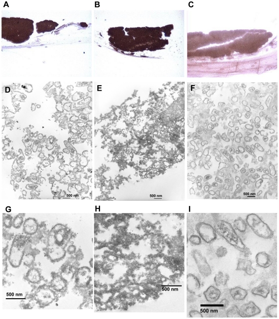
Figure 3. Internalization of IL-1β by A. actinomycetemcomitans co-cultured with organotypic oral mucosa.
Formalin fixed paraffin sections of A. actinomycetemcomitans biofilm containing co-cultures were treated with anti-IL-1β (Panel A), anti-N-terminal-RcpA (Panel B), or control IgG (Panel C) after which the binding antibodies were detected with the NovoLink™ Polymer Detection System (Novocastra™). The sections with the DAB-label were stained with osmium for electron microscopy. Anti-IL-1β stained samples showed structures of A. actinomycetemcomitans cell shape and size (Panel D), with dark precipitate in both extra- and intracellular space (Panel G). The anti-RcpA stained positive control showed intense staining (Panel B) with similar structures (Panel E), although the cell structures are less visible due to the extent of the extracellular precipitate (Panel H). Control IgG antibody showed less staining (Panel C) revealing similar structures (Panel F) without dark precipitates bound to the cell membranes (Panel I).

