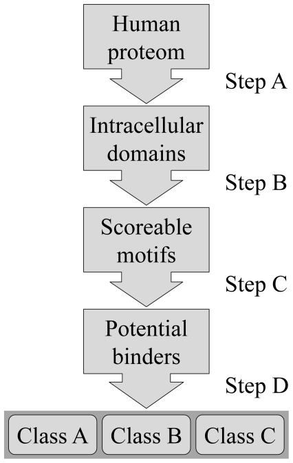
Figure 5. Flowchart of the binding partner prediction.
Step A: all non intracellular segments were excluded from the search. Step B: sequences were split into overlapping eight residue segments; only disordered segments with Gln at position 0 were scored. Step C: a score was assigned (see Methods). Motifs with scores above threshold are considered potential DYNLL binders. Step D: based on amino acid composition, motifs were sorted into three classes with different binding probabilities.

