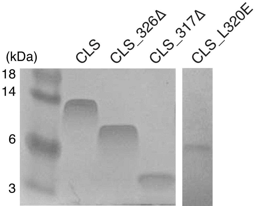
Figure 7. Truncations or mutation of the juxtamembrane region of CLS abolished its slow electrophoretic rate in SDS-PAGE.
Equal amount (5 µg) of purified CLS proteins were loaded onto 15% Tris-glycine SDS polyacrylamide gel. After electrophoresis, the protein bands were stained by colloidal Coomassie blue. Molecular weight markers are shown on the left labeled with corresponding sizes in kDa.

