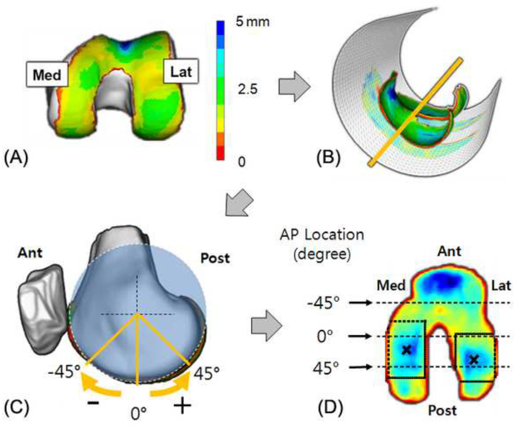
Fig 2.
(a) Femoral cartilage thickness map, (b) a fitted cylinder to project the thickness map, (c) rotational AP location represented as degree relative to the long axis of the femur, (d) rectangular search region on each condyle to find the location of thickest cartilage in the flattened cartilage thickness map

