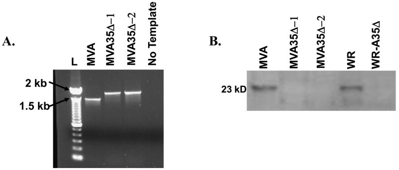
Figure 1. Molecular characterization of MVA35Δ.
a) PCR. MVA-infected cells were transfected with a recombinant PCR fragment containing the E. coli gpt gene inserted between the A35 flanking regions and recombinant viruses were selected in mycophenolic acid-containing media. Virus crude stocks were PCR analyzed using primers in the A35 flanking regions. Wild type A35 locus yields a product of 1400 kbp size and the mutants with gpt inserted yield a size of approx 1900 kbp. L=ladder, b) Western blot showing that A35 is not expressed in MVA35Δ-infected cells. BHK-21 cells were infected with listed viruses at an MOI of 20 for 2 h and analyzed by SDS-PAGE. Blots were incubated with rabbit anti-A35 antibody at a 1:1000 dilution and developed with an anti-rabbit alkaline phosphatase-conjugated secondary antibody.

