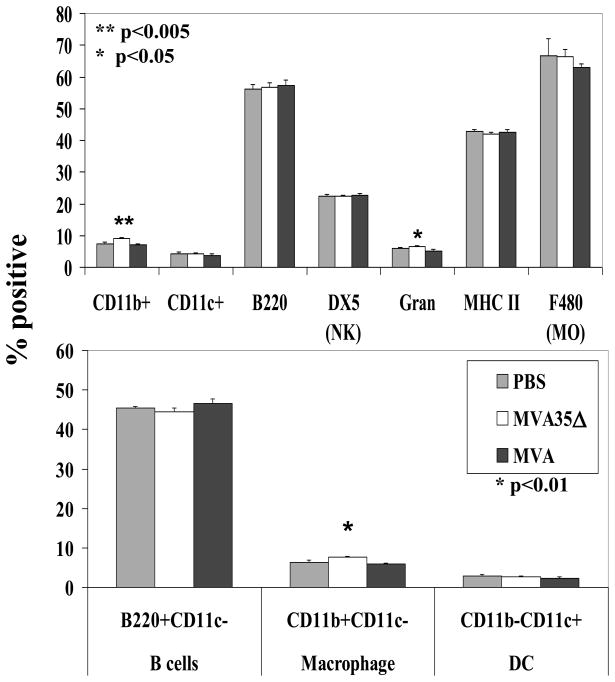
Figure 7. Cellular subsets in spleens.
On day 6 pi, spleens from MVA and MVA35Δ-infected mice (n=5) were stained for various cell surface markers to enumerate percentage of different cell types. Data show average percentage (+/− SEM). Gran, granulocytes; NK, natural killer; MO, macrophage; DC, dendritic cell.

