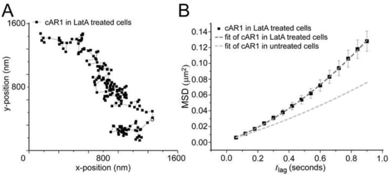
Figure 2. Disruption of actin cytoskeleton increases cAR1 mobility.

(A) Trajectory of a cAR1 molecule after 10 min incubation with 5 μM Latrunculin (n=20 cells). (B) After Latrunculin treatment there were two populations of cAR1 molecules and the fraction sizes were not changed, 30% immobile and 70% mobile. The mobile population still showed directed movement, but the speed of movement was increased D=0.017 ± 0.002μm2/s and a velocity of v= 0.28 ± 0.03 μm/s (n=21 trajectories)
