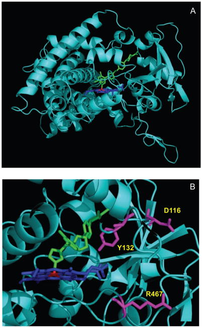
Fig. 7.
Positions of the mutated amino acid residues in CYP51F1. (A) Ribbon diagram of the C. albicans CYP51F1 model using the X-ray crystal structure of M. tuberculosis CYP51 (PDB 1EA1) as a template; (B) The positions of the Y132, D116, and R476 mutations are indicated with colors. Heme and fluconazole are shown in blue and green, respectively.

