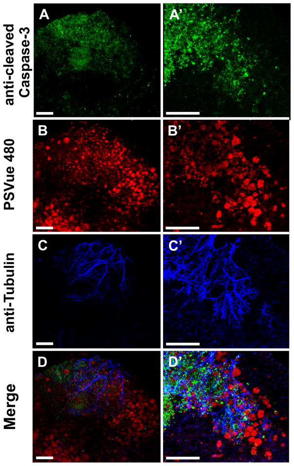
Fig. 7.
Immunochemistry of a Foxg1-cre::Dicer1f/f CKO reveals numerous anti-cleaved caspase 3 positive cells in the forebrain at E12.5 (A, A′). These anti-cleaved caspase 3 positive cells form a gradient with the highest concentration near the rostral (top) pole of the forebrain. Higher power images (A′) indicate that nearly every cell in the most rostral area shows anti-caspase 3 staining. PSVue staining (B, B′) shows prominently stained aggregates of cells in the more caudal and medial parts of the forebrain (B). Higher magnifications reveal surprisingly large aggregates of PSVue-positive cells that match the distribution of opaque cells seen at the macroscopic level (B′). Staining for the neuronal marker anti-acetylated β-tubulin shows fibers most prominent in areas of anti-cleaved caspase 3 staining (C, C′, D, D′) whereas areas of prominent PSVue staining reveal little nerve fiber staining (B, B′, D, D′). This suggests that loss of tubulin correlates with the appearance of phosphatidylserine on the surface of dying neurons. Bar indicates 100 μm.

