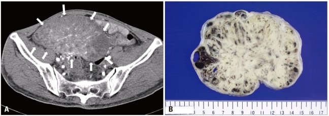
Fig. 3.
Computed tomographic findings of carcinoid tumor. A large mass with inhomogenous density occupies the right pelvic cavity. The mass (white arrow) consists of multiple thick-enhancing septa and enhancing central soft tissue density (A). The right ovary is grossly enlarged; it is measured to be 80 × 124 × 78 mm in size and to weigh 461 g. The ovarian surface is smooth. The cut surface of the mass is bright yellow and solid, and it shows several foci of hemorrhage (B).

