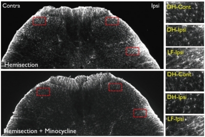
Fig. 8.
GFAP immunoreactivity in the L5 level spinal cord dorsal horn in the Hemi and Hemi+Mino groups. The dotted areas from the low power views are magnified in the right column. When compared with the contralateral side, an increase in GFAP-immunoreactivity is observed in the ipsilateral lateral funiculus (LF-ipsi), but not the ipsilateral dorsal horn (DH-ipsi). In the spinal cord tissue from the Hemi+Mino group, this lateral funiculus-specific increase in GFAP immunoreactivity is attenuated, when compared with the Hemi group.

