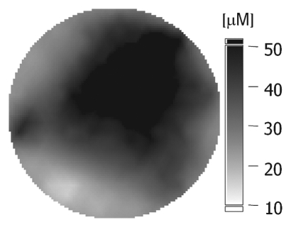
Figure 1b:
(a–c) Imaging and (d) pathologic findings before and after neoadjuvant chemotherapy in 52-year-old woman with pCR. (a) Prechemotherapy axial MR image shows extensive (10.00-cm) clumped enhancement in the upper half of the left breast (arrows) that approximated the distribution of calcifications seen at mammography. An enlarged (2.7-cm) globular left axillary lymph node, with loss of the fatty hilum, indicating metastatic involvement, was also identified at MR imaging but is not shown. (b) Prechemotherapy coronal DOS tomographic image of HbT level shows a maximum value of 45 μmol/L in the tumor ROI. (c) Postchemotherapy coronal DOS tomographic image shows that the HbT level had decreased to background levels in the tumor ROI; this correlated with resolution of both the regional clumped enhancement (primary breast carcinoma) and resolution of the axillary nodal metastasis. (d) Postchemotherapy immunohistochemical slide shows only rare tumor-induced CD105 (endoglin)-expressing blood vessels (arrow) in the area of the prior tumor. These findings correlated with resolution of both the regional clumped enhancement (primary breast carcinoma) and resolution of the axillary nodal metastasis. (Diaminobenzidine-detection substrate, hematoxylin counterstain; original magnification, ×200.) Histopathologic findings in the mastectomy specimen in this patient are shown in Figure E1 (online).

