
Figure 2d:
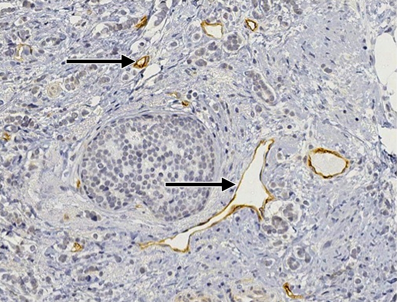
(a–c) Imaging and (d) pathologic findings before and after neoadjuvant chemotherapy in 51-year-old woman with pPR. (a) Prechemotherapy axial MR image shows an irregularly shaped enhancing mass with spiculated margins that measures 5.8 cm in its greatest transverse diameter (between arrows). (b) Prechemotherapy coronal DOS tomographic image of HbT level shows a maximum value of 50 μmol/L in the tumor ROI. (c) Postchemotherapy coronal DOS tomographic image shows that HbT remained at the same level in the tumor ROI and correlated with reduced but persistent malignant enhancement of the residual tumor. (d) Postchemotherapy immunohistochemical slide shows abundant tumor-induced CD105 (endoglin)-expressing blood vessels (arrows) in areas associated with residual tumor. (Diaminobenzidine-detection substrate, hematoxylin counterstain; original magnification, ×200.) Histopathologic findings in the mastectomy specimen in this patient are shown in Figure E2 (online).