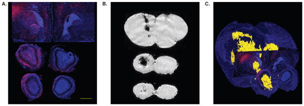
Figure 1. High-resolution ex vivo MR imaging of neonate shiverer mouse brain following intracerebroventricular transplantation of superparamagnetic ion oxide-labeled immortalized neural stem cells.
The mismatch between conventional histology and loss of MR detectability following cell proliferation is apparent. (A) Two weeks after grafting, Feridex-labeled C17.2 cells migrated vast distances toward the outer cortical layers of the cerebrum and olfactory bulb as revealed by anti-β-gal staining. This is in sharp contrast with the MR imaging pattern (B), which shows hypointense cells centered in and around the ventricles, but not the cortical layers. The merged histology/MR image (C), in which β-gal+ cells are red, and MRI hypointense cells yellow, further illustrates this mismatch. Scale bar = 1 mm.
Reproduced, with permission, from Walczak et al. [13].

