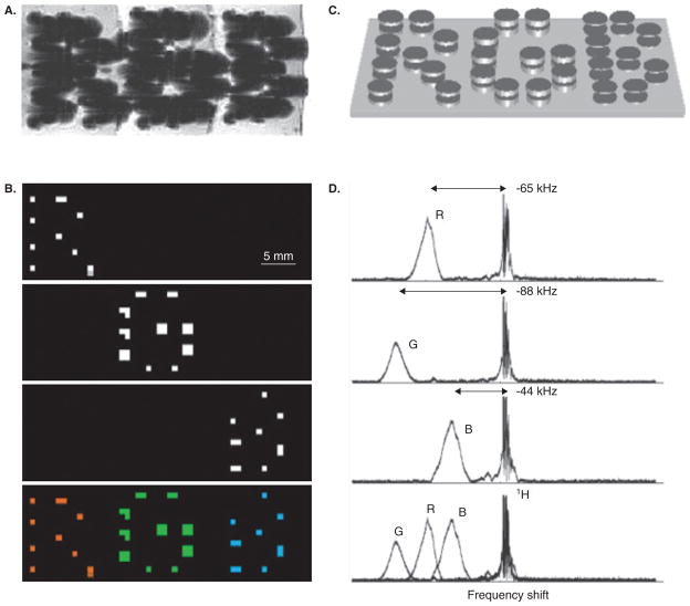
Figure 4. Micro-engineered magnetic particles for multi-color MR imaging.
The particle frequency was varied by changing the thickness of electroplated nickel layers that formed the magnetizable disk pairs. (A) As with normal superparamagnetic ion oxide detection, magnetic dephasing due to the particles’ external fields enables the spatial imaging shown in the gradient-echo MRI. However, comparison between (A) and the chemical shift images (B) shows that the additional spectral information both differentiates between particle types and improves particle localization. The particles are shown schematically (not to scale) in (C). With particle spectra [(D), to the right of the corresponding chemical shift images in (B)] shifted well clear of the water proton line, different planes in the chemical shift imaging map isolate different particle types for unambiguous colour-coding with minimal background interference (B, bottom panel).
Reproduced, with permission from Zabow et al. [40].

