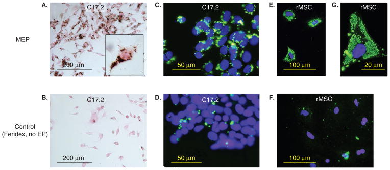
Figure 5. Superparamagnetic ion oxide (SPIO)-labeling of cells using magnetoelectroporation.
C17.2 mouse neural stem cells (A – D) and rat mesenchymal stem cells (rMSC) (E – G) were incubated with Feridex (2 mg Fe/ml) with (A, C, E, G) and without (B, D, F) electroporation (EP). Only MEP-treated cells show significant Feridex uptake as assessed by DAB-enhanced Prussian blue stain (A) and anti-dextran immunofluorescence (C, E). The higher magnification in (G) demonstrates Feridex-containing clusters with a measured diameter of 830 ± 350 nm.
Reproduced, with permission, from Walczak et al. [53].

