
Figure 6. In vivo MRI of magnetically labeled dendritic cells used as cancer vaccines in melanoma patients.
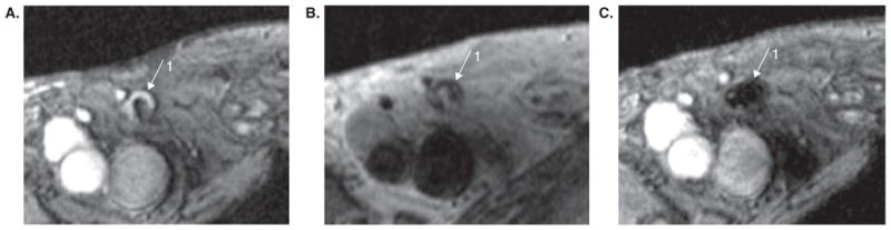
(A) Gradient echo MR image before vaccination showing inguinal lymph node as a hyperintense signal area (1). (B) Spin echo MR image showing same lymph node after vaccination. (C) Gradient echo (technique more sensitive to superparamagnetic ion oxide) MR image after vaccination in same position as (B) showing a marked decrease in signal intensity from lymph node 1.
Reproduced, with permission, from de Vries et al. [60].