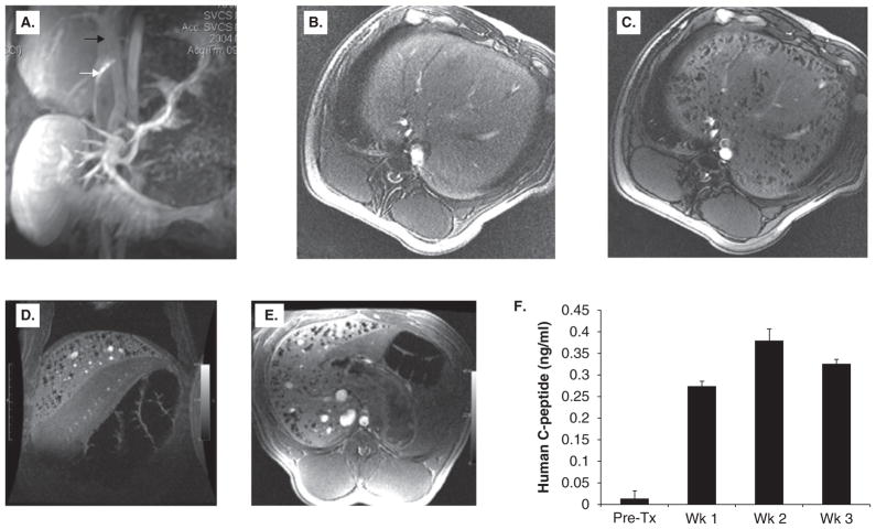
Figure 7. MR-guided injection of magnetocapsules in swine.
(A) Conventional magnetic resonance angiography/venography of the mesenteric venous system was performed with Gadolinium- diethylene-triamine-pentaacetic acid before any punctures. White arrow, active needle; black arrow, portal vein. The needle is seen in the inferior vena cava in the proper orientation for porto-caval puncture. (B,C) In vivo MRI of magnetocapsules before (B) and 5 min after (C) intraportal infusion of magnetocapsules in a pig. Magnetocapsules can be seen distributed throughout the liver as hypointense signal voids created by superparamagnetic ion oxide particles. (D,E) MRI follow up at three weeks shows the persistence of magnetocapsules as intact signal voids. (F) Magneto-encapsulated human islets retain functionality in vivo, as assessed by a sustained increase in human C-peptide in plasma.
Reproduced, with permission, from Barnett et al. [97]

