
Figure 1.
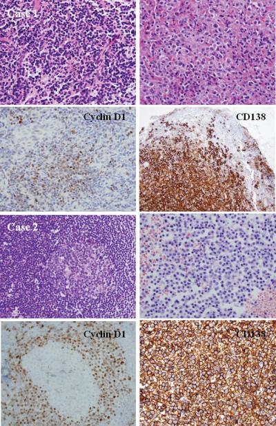
A. Histologic and phenotypic features of MCL with clonal PC differentiation (case 1): a naso-pharyngeal mass showing a diffuse pattern of atypical lymphoid cells. Clusters of PCs are present and adjacent to the lymphoid aggregates (H&E stain, upper left and right, original magnification 100X). Immunohistochemical staining for Cyclin D1 and CD138 shows the MCL and PC component, respectively (immunoperoxidase stain, lower left and right, original magnification 100X). The two clones were unrelated. This case might represent a concomitant plasma cell neoplasm or a marginal zone lymphoma and MCL in the same patient. B. Histologic and phenotypic features of MCL with clonal PC differentiation (case 2) in a lymph node showing reactive GC (upper left) with expanded mantle zone. Clusters of PCs are seen in the bone marrow biopsy (H&E stain, upper left and right, original magnification 100X). Cyclin D1 stain shows a mantle-zone growth pattern, whereas CD138 stain shows numerous positive PCs (immunoperoxidase stains, original magnification 100X). The two populations were clonally related, and both harbored the t(11;14)(q13;q32).