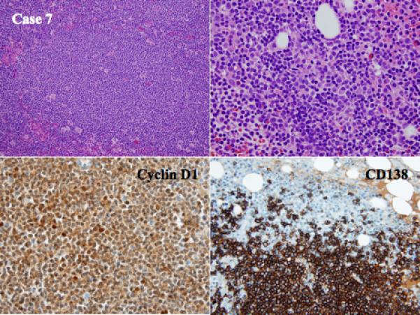
Figure 4.

Histologic and phenotypic features of MCL with clonal PC differentiation (case 7): a combined nodular/diffuse pattern of MCL in a lymph node and bone marrow biopsy of the same patient showing an admixture of monotypic PCs and MCL aggregates (H&E stain, upper left and right, original magnification 40X and 100X). Immunohistochemical staining for Cyclin D1 and CD138 shows the MCL and PC component, respectively (immunoperoxidase stain, lower left and right, original magnification 100X). The two neoplastic populations are derived from the same clone.
