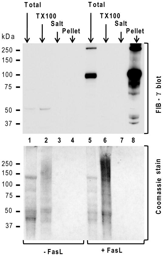
Figure 3. Solubility dynamics of FIB-γ during FasL-induced liver injury.
Solubility dynamics of FIB-γ during FasL-induced liver injury were analyzed biochemically by comparing the soluble TX100 fraction, high salt wash fraction (Salt) and the insoluble fraction (Pellet) from FasL-treated and untreated livers. TLL (Total) were used as controls. None of the fractions showed detectable levels of FIB-γ in untreated liver samples. However, FIB-γ antibody faintly recognized the FIB-γ monomer (50-kDa) in the TLL and the soluble TX100 fraction of untreated liver. In contrast, FIB-γ blot showed two major bands (100-kDa, 250-kDa) in FasL-treated TLL, but not in the soluble TX100 and wash fractions. A duplicate gel to that used for immunoblotting was stained with Coomassie blue. Athough the protein amounts in some of the lanes (3,4,7,8) were not enough to be visible with Coomassie staining, they were readily detected by the FIB-γ antibody (lane 8).

