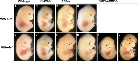
Figure 2.
Gross morphology of embryos from dams maintained on vitamin A-sufficient (VitA-suff) dietary regimen (A, C, E, G) or vitamin A-deficient (VitA-def) dietary regimen (B, D, F, H–J) during pregnancy. Wild-type (A, B), CMOI−/− (C, D), RBP−/− (E, F), and CMOI−/−RBP−/− (G–J) embryos from dams of the same genotype, respectively, were collected at 14.5 dpc. e, abnormal eye (reduced pigmentation in the ventral region); p, peripheral edema; mf, abnormal midfacial region (snout foreshortened and divided by a sagittal median cleft, prolabium absent, maxillary process bearing whiskers separated by a larger than normal distance); ex, exencephaly (exteriorized brain). Same magnification was used for all panels. Panels A, B, E, F were previously published (23).

