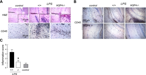
Figure 5.
Reduced brain inflammation in AQP4-null mice after intracerebral LPS injection. Mice were injected intracerebrally with PBS (control) or LPS. A) Brain histology at 1 d by H&E staining (top panels) and CD45 immunocytochemistry (bottom panels). B) Gallery of CD45-immunostained sections of hippocampus from 3 wild-type and 3 AQP4-null mice. C) Quantification of inflammation done using H&E and CD45-stained sections (se). *P < 0.01 vs. +/+.

