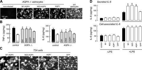
Figure 8.
Evidence for aquaporin-facilitated cytokine secretion. A) Immunofluorescence of AQP4 (M1 and M23 isoforms) and AQP1 in adenovirus-treated primary astrocyte cultures from AQP4-null mice (AQP4−/− astrocytes). AQP4 immunofluorescence of culture from wild-type mice shown at right. B) TNF-α and IL-6 in culture medium at 2 h after LPS (100 ng/ml) or saline addition (se, n=4). Representative of 3 sets of cultures/infections. *P < 0.01 vs. −/− control. C) AQP4 and AQP1 immunofluorescence, and GFP fluorescence, of adenovirus-treated T24 (bladder) cells. D) Secreted IL-6 (in culture medium, top panel) and cell-associated IL-6 (bottom panel) at 2 h after addition of LPS (se, n=4). *P < 0.01 vs. LPS-treated control.

