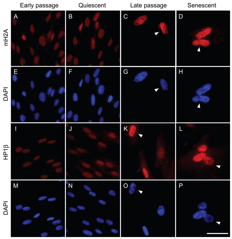
Fig. 1.
Representative immunofluorescent images of mH2A and HP1β protein expression in the nuclei of human fibroblasts at different replicative ages. LF1 cells were stained and images acquired as described in Experimental procedures. The mH2A antibody was directly conjugated to Alexa Fluor 647. HP1β was visualized using a secondary antibody conjugated to Cy3. Early passage: vigorously growing cultures within the first third of their replicative life span. Quiescent: early passage cells were contact inhibited and then cultured in 0.25% serum for 48 hr. Late passage: cultures within 8–12 population doublings of their terminal senescent arrest. Senescent: cultures whose cell numbers have not increased for a minimum of 2 weeks. Flow cytometric cell cycle profiles for early passage, quiescent and senescent cultures are shown in Fig. S2. Arrowheads indicate the variety of SAHF morphologies seen in LF1 cells: cell showing robust mH2A and DAPI foci, panels D,H; cell showing intermediate but poorly resolved HP1β and DAPI foci, panels K,O; cell showing very few and small HP1β foci and no DAPI foci, panels L,P; cell showing no mH2A or DAPI foci but high overall levels of mH2A, panels C,G.

