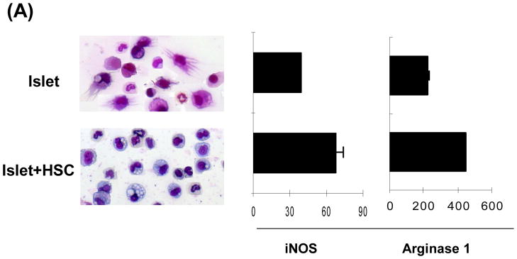
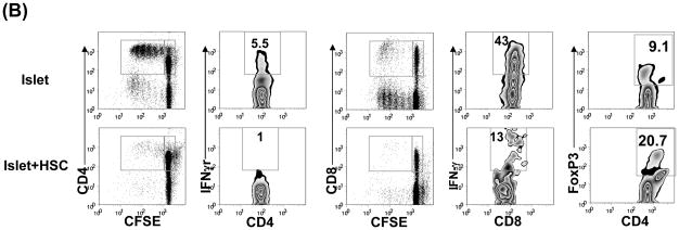
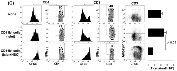
Figure 2. Characterization of CD11b+ cells isolated from islet/HSC grafts.
CD11b+ cells were purified by magnetic beads from cells isolated from BALB/c (H-2d) islet allografts cotransplanted with B6 (H-2b) HSC on POD 7. Islet alone allografts served for comparison (n=3 in each group). (A) Cell morphology (Giemsa staining) and expression of iNOS and arginase 1 [quantitative (q) PCR]. The data are expressed as mean relative to 18S ± 1SD. (B) Allo-stimulatory activity of the isolated CD11b+ cells. Irradiated CD11b+ cells pulsed with BALB/c spleen cell lysate (without alloantigen pulsing served as control), were cultured with CFSE labeled B6 spleen T cells at a 1:20 ratio for 5 days. Proliferative responses were determined by CFSE dilution. Expression of IFN-γ and Foxp3 was assayed by intracellular staining with specific mAbs, and analyzed by flow cytometry. The number is percentage of positive cells in CD4 or CD8 T cell subset. (C) CD11b+ cells from islet/HSC grafts inhibit T cell response. CD11b+ cells isolated from islet alone or islet/HSC grafts were added at the beginning into the culture of CFSE-labeled T cells, proliferation of which was elicited by anti-CD3 mAb (2 μg/ml) for 3 days. Without addition of CD11b+ cells served as control. Expression of IFN-γ was measured by intracellular staining with specific mAbs. Apoptotic activity was measured by staining with anti-annexin V mAb. The number is percentage of positive cells in T cells or their subsets. CD3+ cells in each well was counted, and expressed as mean ± 1SD.


