
Figure 7. H-MC inhibit T cell response in vivo.
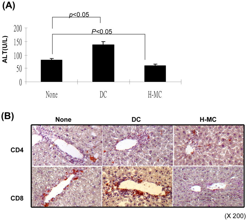

(A) Administration of H-MC attenuates liver damage. DC or H-MC (1.5 × 10⁶) that had been pulsed with OVA were intravenously injected into OVA-HEP mice whose liver injury was induced by adoptive transfer of T cells from OT-1 (5 × 10⁶) and OT-II (2 × 10⁶) mice. Serum was obtained on day 3 post cell injection, and tested for ALT with specific kit. (B) Administration of H-MC leads to less antigen specific CD8+ infiltrating cells in the liver. The animals were sacrificed on day 6 post cell transfer. The liver cryostat sections were histochemically stained with anti-CD4 or -CD8 mAb, and examined by microscopy. (C) A total of 30 high power fields (hpf) were randomly selected in each liver (n=3), and counted for CD4+ and CD8+ cells. The data are expressed as cell number/hpf ± 1SD. (D) Cotransplantation with H-MC effectively protects islet allografts. 300 BALB/c islets were mixed with 2, 5 × 10⁶ of H-MC or DC generated from B6 BM by co-culture with HSC either from B6, BALB/c or C3H strain, and transplanted into diabetic B6 recipients. Islet allografts alone served as controls. The islet graft survival was monitored by blood glucose level as described in Methods.