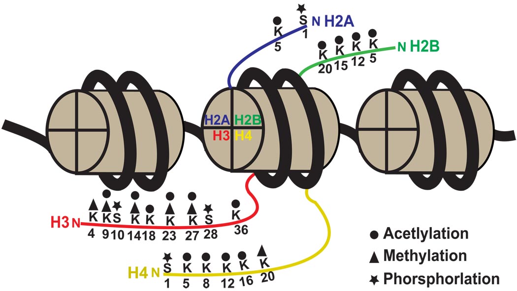
Figure 1. Schematic illustration of the nucleosome structure.
The nucleosome contains 147 base pairs of DNA (bold black line) that encircle an octameric core of histone proteins. Three nucleosomes are illustrated. The histone octamer is composed of two molecules of each of the canonical histone 2A (blue), 2B (green), 3 (red) and 4 (yellow) proteins. The N-terminal tails (colored thin lines) of these histone proteins are subject to a variety of covalent modifications, including acetylation, methylation and phosphorylation. Numbers beneath the colored lines denote the position of amino acid residues of the histones that are covalently modified. Letters above the colored lines indicate the amino acid substrates for these modifications. K: lysine, S: serine.

