Fig. 4.
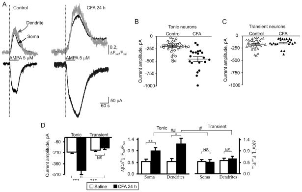
AMPAR-mediated currents and associated [Ca2+]i transients are markedly potentiated in tonic but not in transient SG neurons during persistent inflammation. (A) Representative examples of AMPA-induced currents (lower traces) and [Ca2+]i transients (upper traces) in soma (black traces) and dendrites (grey traces) in tonic neurons 24 h after saline (control) or CFA. (B, C) Scatter dot plots illustrate a spread in extrasynaptic AMPAR-mediated currents in tonic (B) and transient (C) neurons 24 h after saline or CFA. (D) A statistical summary of current amplitudes (left graph) and [Ca2+]i transients (right two graphs) in soma and dendrites of different groups of SG neurons 24 h post-saline and post-CFA. * p < 0.05, ** p < 0.001, *** p < 0.0001 versus the saline-treated group; # p < 0.05, ## p < 0.001 versus the transient SG neurons; NS, not significant.

