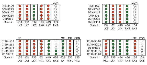
Figure 3. Detailed LOH analysis for non-targeted chromosomes.
LOH events for chromosomes 6, 7, 12, or 14, detected in the genome-wide screen shown in Figure 2, were examined with additional polymorphic loci for these chromosomes. Unless indicated otherwise (MR, mitotic recombination; IE, intragenic event; CON, control; see Figure 1), the Aprt mutant clones examined exhibited loss of chromosome 8. Control clones (CON) were chosen from those that did not exhibit an LOH event for the chromosome examined in the initial screen (Figure 2) though examples of MR were detected in some controls (see mutant 476RK2, chromosome 12 analysis for example). “Clone #” indicates mouse tag number, left or right kidney (LK or RK) and clone number. Red circle indicates loss of a DBA/2 locus and green circle indicates loss of C57BL/6 locus. Open circles indicate retention of heterozygosity.

