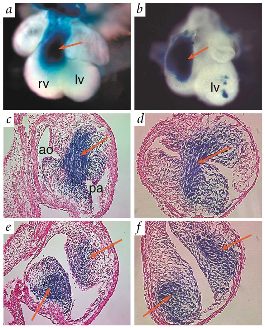
Fig. 4.
Fate mapping of cardiac neural crest. a,b, Hearts of E12.5 wild-type (a) and Nf1−/− (b) embryos stained for β-galactosidase activity. Cardiac neural-crest cells are labeled blue by Cre-mediated activation of lacZ in R26R reporter mice30. Red arrows point to labeled cells populating the outflow tract of the developing heart. c,d, Cross-sections through hearts at the level of the truncus show labeled blue cells forming the septum between the aorta and pulmonary artery in both wild-type (c) and Nf1−/− (d) embryos. e,f, Red arrows point to labeled neural-crest cells invading the outflow tract endocardial cushions in the conus region of the right ventricular outflow tract in wild-type (e) and Nf1−/− (f) embryos. pa, pulmonary artery; ao, aorta; rv, right ventricle; lv, left ventricle.

