Figure 5.
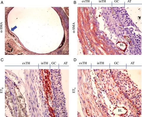
Localization of αSMA, ETA and ETB protein in large antral follicle. Specific antibodies for αSMA (A and B), ETB (C) and ETA (D) proteins were used to stain adjacent sections of a normal human ovary followed by counter staining by Mayer's hematoxylin. The high magnification images (×400; B–D) were taken from the marked (arrow) area of (A) (×12.5). inTH, theca interna; exTH, theca externa; BL, blood vessel; AT, antrum. Note that ETB expression is specific to theca interna (C), whereas ETA expression is seen in the GC, theca interna and theca externa (D) where αSMA staining is most intense (B). No positive signal was seen in the negative controls (no primary antibody; not shown). These are representative images from one of the seven ovaries that had at least one healthy large follicle in each ovary.

