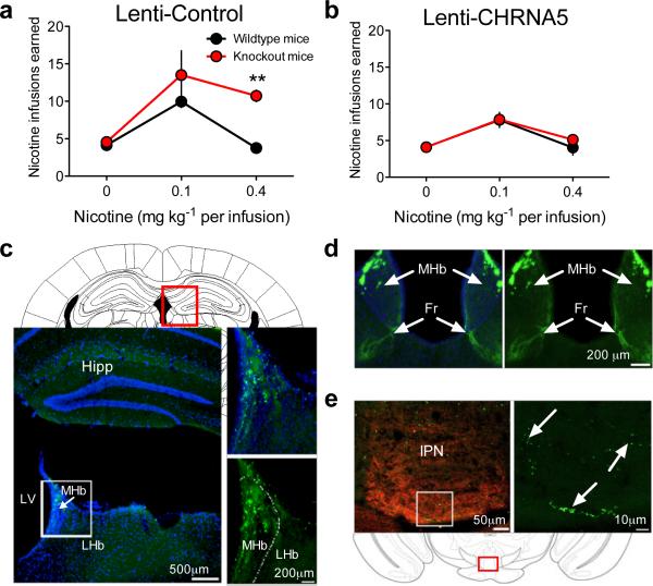
Figure 2. “Rescue” of α5* nAChRs in MHb-IPN normalizes nicotine intake.
(a) Mean (± SEM) nicotine infusions in Lenti-Control mice. Genotype F(1,22)=7.70, p<0.05; Dose F(2,22)=19.34, p<0.0001; Interaction F(2,22)=3.75, p<0.05. **P<0.01 between genotypes (b) (± SEM) nicotine infusions in Lenti-CHRNA5 mice. Genotype F(1.28)=0.17, not significant (n.s.); Dose F(2,28)=16.05, p<0.0001; Interaction F(2,28)=0.36, n.s.; n=6–9 per group. (c) GFP Immunostaining confirmed MHB virus delivery. Hipp, hippocampus; LHB, lateral habenula; LV lateral ventricle; MHb, medial habenula. (d) GFP-labeled cells in MHb, DAPI-counterstained in left panel, extend into the fasciculus retroflexus (Fr). (e) GFP-positive axons detected in IPN. Left panel is labeled with VAChT (red) to identify IPN.

