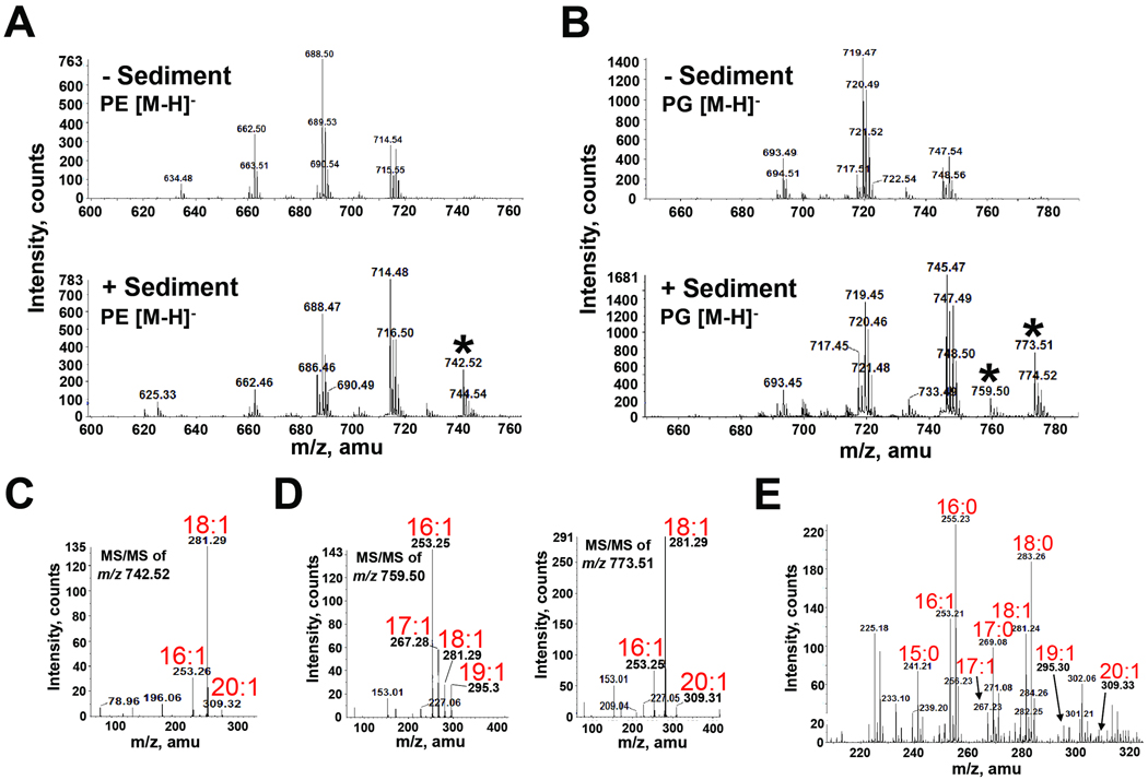
Fig. 9. Mass spectrometric analysis of PG and PE extracted from V. cholerae grown in the presence and absence of sediment indicates assimilation of fatty acids present in ocean sediment.
A and B. Negative-ion ESI mass spectra of [M-H]− ions of V. cholerae PE (A) and PG (B) isolated from cultures grown in the presence and absence of sediment. Selected peaks (*) found only in the presence of sediment were subsequently analyzed by MS/MS.
C. Negative-ion MS/MS of the PE [M-H]− ion at m/z 742.52 detected only in the presence of sediment. This product-ion spectra revealed the presence of the monounsaturated fatty acid 20:1 as a constituent of PE.
D. Negative-ion MS/MS analysis of the product-ion spectra of the PG [M-H]− ions at m/z 759.5 and 773.51 detected only in the presence of sediment. These [M-H]− ions revealed the presence of the monounsaturated fatty acids 17:1, 19:1 and 20:1 as constituents of PG.
E. Negative-ion ESI-MS analysis of the fatty acid content in sediment identified unique fatty acids (17:1, 19:1 and 20:1) found in V. cholerae PLs following growth in sediment.

