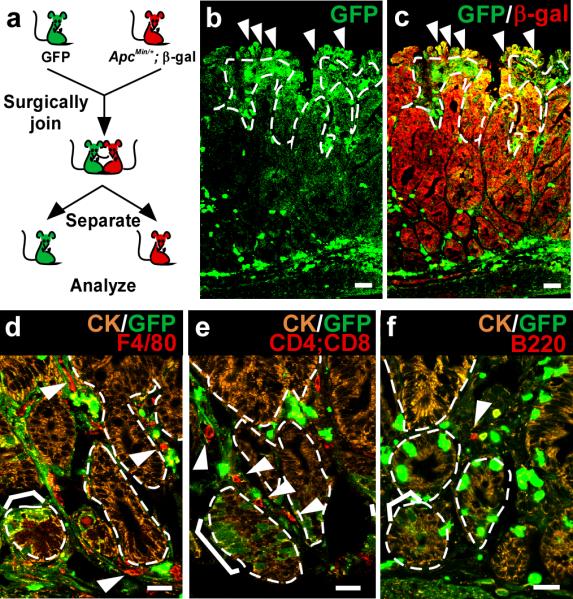
Figure 1. Intestinal cell fusion in tumorigenesis.

a, Parabiosis experimental design. GFP and ApcMin/+;ROSA26 mice were surgically joined. b-c, Cell fusion was observed in small intestinal polyps. Single plane confocal microscopy images of GFP (green) and β-galactosidase (red) detected by antibodies demonstrate fusion by co-localization in yellow (c). Arrowheads denote examples of fused cells. d-f, Lymphocytes and leukocytes are present within small intestinal polyps that have undergone fusion. Single plane confocal microscopy depicts fusion-derived (brackets) and unfused epithelia detected with antibodies to GFP (green) and cytokeratin (marking the epithelial compartment, orange). Arrowheads indicate F4/80+ macrophages (red) (d), CD4+ or CD8+ T cells (red) (e), or B220+ B cells (red) (f) in the tumor mesenchyme. Dashed white lines indicate epithelial/mesenchymal border. Bars=25μm.
