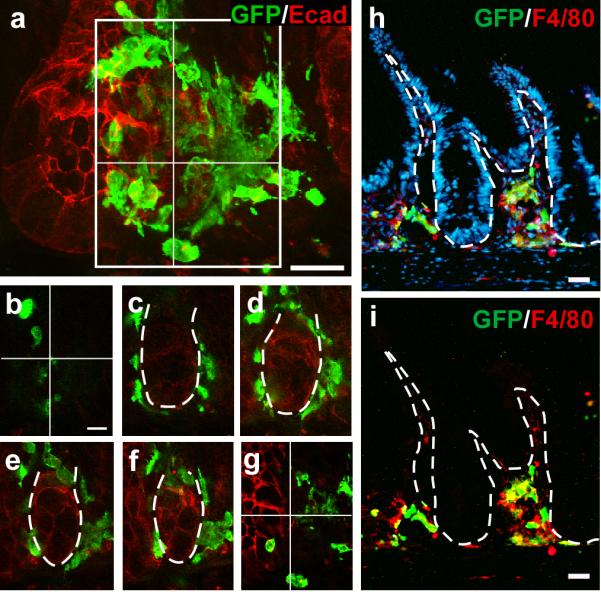
Figure 3. Pre-fusion clusters of BMDCs contain macrophages.

a, 50μm tissue section and 3-dimensional crypt reconstruction of a confocal Z-stack image illustrating GFP-positive BMDCs (green) surrounding the crypt/stem cell niche. Epithelial cells are marked with E-cadherin (red). b-g, Sequential sections through the Z-stack shown in (a). h-i, Pre-fusion cell clusters contain F4/80-positive macrophages (red) consisting of both donor-derived (yellow) and non-donor-derived cells. Nuclei are stained with Hoechst (h; blue). Dashed white lines indicate epithelial/mesenchymal border. Bars=25μm.
