
Figure 2.
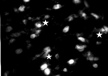
Representative fluorescent image of cardiomyocytes treated with ceramide during cardioplegia (60 min) and reperfusion (20 min) (group III). After DAPI counterstaining the greater nuclei of cardiomyocytes allow their distinction from fibroblasts with smaller nuclei. In anti-activated caspase-3 positive, apoptotic cardiomyocytes the cytoplasm reveales an intensive granular fluorescence (marked with stars). The exemplary images represent a single experiment. During the cryosection procedure artifacts presenting as nuclei conglomerates could not be avoided; these were excluded from analyses.